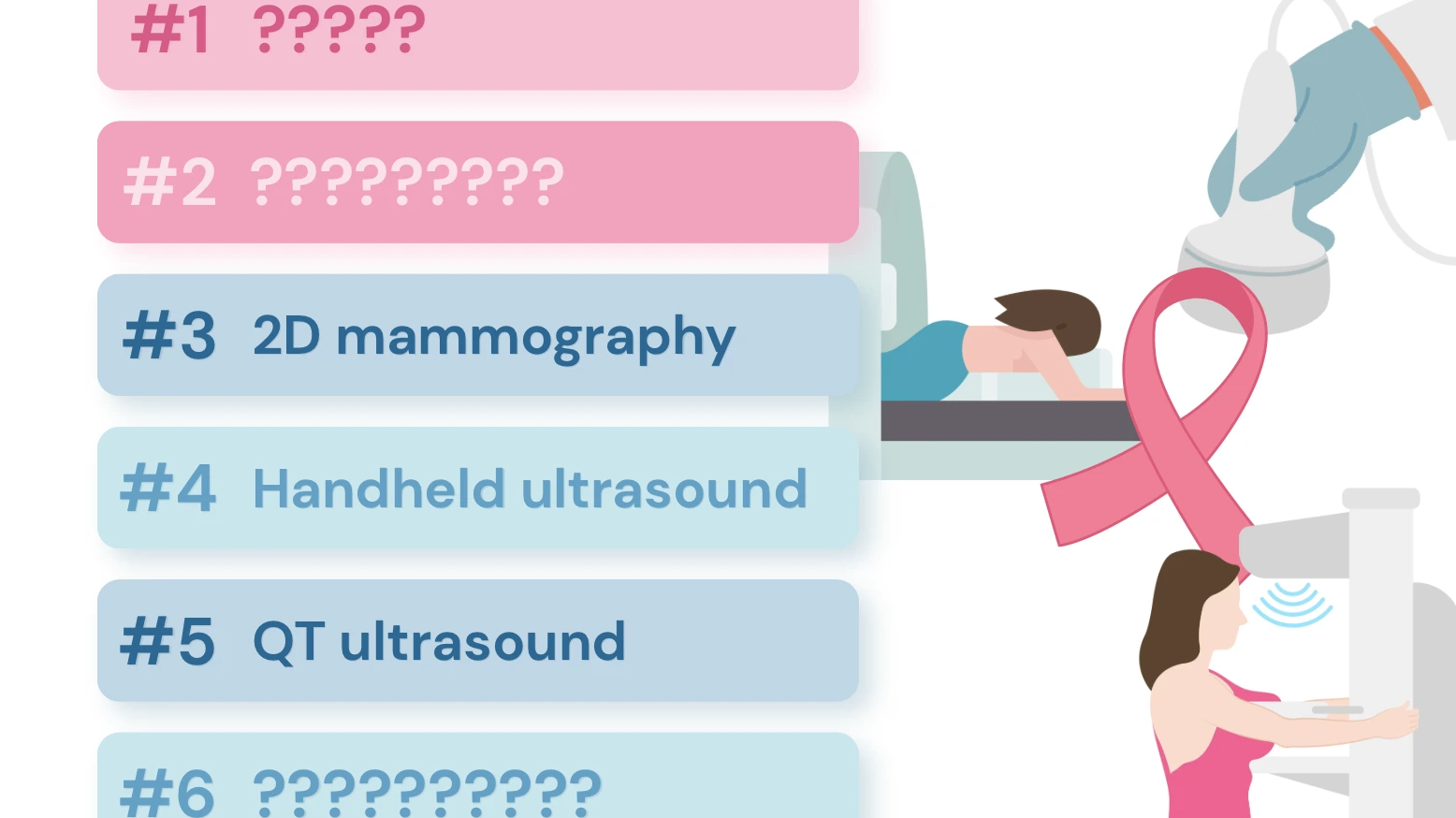
We ranked six breast cancer screening tools – 2D and 3D mammography, breast MRI, ultrasound, QT ultrasound, and whole-body MRI – by what the research shows.
On a mammogram, both cancer and dense breast tissue show up white. Radiologists call it looking for a snowball in a snowstorm. Nearly half of women over 40 have dense breasts, and for them, screening and early detection is an uphill battle.
This Breast Cancer Awareness Month, we're doing something most screening guides don't: putting every major breast imaging option side by side and ranking them honestly – including the ones that are newer, flashier, and even the ones that aren't built for this job.
Quick answer: For finding breast cancer, contrast-enhanced breast MRI is the most sensitive test available, followed by 3D mammography (tomosynthesis) and then standard 2D mammography. Handheld ultrasound is a useful add-on for dense breasts.
QT (transmission) ultrasound is promising but early. Whole-body MRI is not a substitute for dedicated breast screening. For most women, the best plan is a mammogram plus the right supplemental test for their risk and breast density – not one test alone.
How We Ranked Them
We ranked each test on one question first: if a cancer is there, how likely is this test to find it? That's sensitivity, and it's measured best by cancer detection rate (cancers found per 1,000 women screened) and by how many cancers slip through between screens (“interval cancers”).
Then we weighed four tiebreakers:
- Strength of evidence. Large randomized trials outrank small single-company studies.
- False alarms. Every extra callback or biopsy has a real cost in anxiety, time, and money.
- Safety and comfort. Radiation, compression, IV contrast, and time in the scanner.
- Real-world access. Guidelines, insurance coverage, and availability.
The Ranking, from #6 to #1
#6: Whole-Body MRI: Excellent at Its Job, but Breast Screening Isn't It
Whole-body MRI surveys the brain, organs, spine, and pelvis in a single session, with no radiation. It can catch things nobody was looking for. But a full-body protocol is not a breast exam.
Dedicated breast MRI uses a specialized breast coil, the patient lying face-down, thin high-resolution slices, and, critically, an IV contrast agent (gadolinium) timed to show how blood flows into tissue. Cancers build their own blood supply, and that pattern of enhancement is what makes breast MRI so sensitive.
Bottom line: Whole-body MRI is a valuable whole-picture health scan. Think of it as a complement to breast screening, never a replacement.
#5: QT (Quantitative Transmission) Ultrasound: The Most Interesting Newcomer
QT ultrasound is genuinely new and different. Instead of a technologist moving a probe, you lie face-down with one breast in a warm water bath while transducers send sound waves through the tissue, building a 3D map. No radiation, no compression, no contrast injection.
The device is FDA-cleared as a breast ultrasound system. In an early multi-reader study by the manufacturer's research team, readers looking at transmission ultrasound images showed modestly higher sensitivity and specificity than with 2D mammography, with fewer callbacks. Speed-of-sound measurements also helped readers tell fluid-filled cysts from solid lumps with over 92% accuracy.
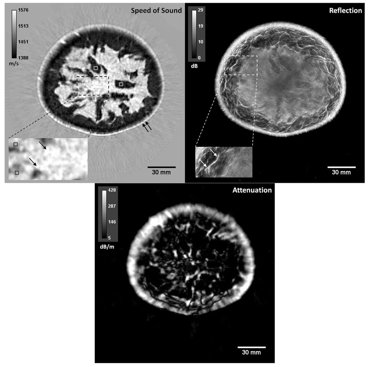
Image 1: Multimodality QT ultrasound images of a breast | From Objective breast tissue image classification using Quantitative Transmission ultrasound tomography 2016
It is of note that that study included 108 women, was retrospective, and was industry-authored. A head-to-head prospective comparison against 3D mammography has not yet been published.
Bottom line: Promising and comfortable, especially for younger women who want to avoid radiation. But not yet proven enough to stand alone.
#4: Handheld (Diagnostic) Ultrasound: The Dense-Breast Sidekick
Ultrasound is the easy first step for a lump you can feel and for a spot flagged on a mammogram with no radiation, no contrast, and only small area compression. As a screening add-on for dense breasts, it also has solid trial evidence.
In the ACRIN 6666 trial of 2,809 women with dense breasts and elevated risk, adding annual ultrasound to mammography found an extra 4.3 cancers per 1,000 women per year. In Japan's J-START randomized trial of more than 72,000 women in their 40s, adding ultrasound raised screening sensitivity from 77.0% to 91.1%.
The trade-off is false alarms. Ultrasound finds many benign lumps that look worrisome, and results depend heavily on the operator's skill. It also misses many of the tiny calcifications that signal early cancers on a mammogram.
Bottom line: A strong, radiation-free supplement when MRI isn't available or appropriate, especially for dense breasts.
#3: 2D Digital Mammography: The Proven Foundation
Standard mammography is the only screening test with decades of randomized-trial evidence showing fewer breast cancer deaths. It is fast, widely covered, and excellent at catching microcalcifications — the grain-of-sand flecks that are often the earliest sign of ductal carcinoma in situ (DCIS).
Its weakness is density. In a study of more than 329,000 screening mammograms, sensitivity ranged from 87.0% in women with mostly fatty breasts to 62.9% in women with extremely dense breasts. That gap is why the FDA now requires every U.S. mammogram report to tell women whether their breasts are dense.
Bottom line: Still the backbone of screening. If 3D isn't available to you, a 2D mammogram is far better than none.
Drumroll for our final two modalities, which tie for first place due to just how different they are and the value they offer.
#1B: 3D Mammography: The Better Everyday Mammogram
Tomosynthesis takes many low-dose X-ray images from different angles and reconstructs the breast in thin slices. Overlapping tissue that might hide a cancer, or mimic one, gets separated.
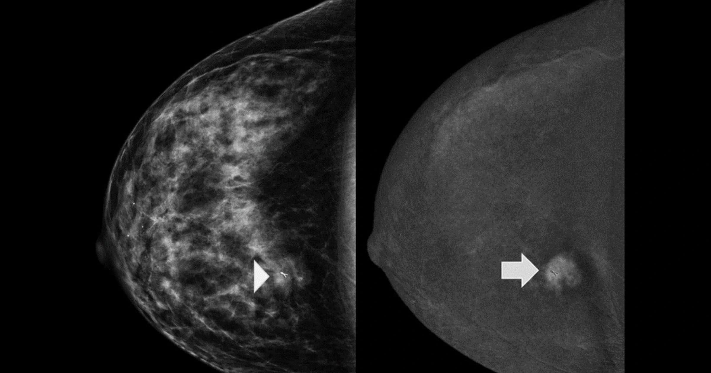
In a study of nearly 455,000 exams across 13 U.S. sites, adding 3D raised cancer detection from 4.2 to 5.4 per 1,000 screens and cut callbacks from 107 to 91 per 1,000. Invasive cancer detection rose by 41%.
The benefit isn't equal for everyone. A large community-practice analysis found 3D helped most in women with scattered or heterogeneously dense tissue, but offered little added cancer detection for women with extremely dense breasts. A large NCI-sponsored randomized trial, TMIST, is still testing whether 3D lowers advanced cancers over time.
Bottom line: The best routine screening mammogram available today. If you have extremely dense breasts, it still may not be enough on its own.
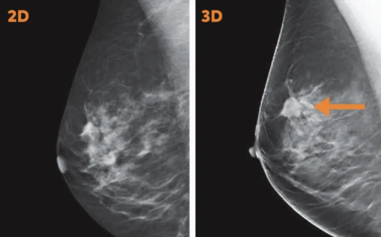
Image 2: 2D vs 3D mammogram | Via Allevia Radiology
#1A: Breast MRI with Contrast: The Most Sensitive Test We Have
Contrast-enhanced breast MRI finds cancer by watching blood flow, not density, so dense tissue barely slows it down. The evidence is consistent across major trials:
- DENSE trial (Netherlands, 40,373 women with extremely dense breasts and normal mammograms): MRI found 16.5 additional cancers per 1,000 women, and cut interval cancers in half, from 5.0 to 2.5 per 1,000 among women invited, and to 0.8 per 1,000 among women who actually had the MRI.
- EA1141 trial (1,444 women with dense breasts): Abbreviated breast MRI found every one of the 17 invasive cancers; 3D mammography found 7. Detection was 11.8 vs. 4.8 per 1,000.
- ACRIN 6666: Even after both mammography and ultrasound came back negative, a single MRI found 14.7 more cancers per 1,000 women.
But with sensitivity comes tradeoffs. It produces more false positives (about 8% in DENSE's first round). It requires an IV gadolinium contrast injection. It can miss some calcification-only DCIS, which is why it is paired with mammography rather than replacing it. And U.S. guidelines are split: the USPSTF (2024) says evidence is insufficient to recommend supplemental MRI for dense breasts, while the American College of Radiology recommends supplemental MRI for women with extremely dense breasts and for those at higher-than-average risk.
Bottom line: When the goal is to find cancer early, nothing outperforms it. TrueScan offers contrast-enhanced breast MRI as a supplement to mammography for women who want the most thorough look available.
Side-by-Side Comparison
| Rank | Test | Best for | Works in dense breasts? | Radiation | Contrast | False alarms | Strength of evidence |
|---|---|---|---|---|---|---|---|
| 1A | Breast MRI with contrast | High risk; dense breasts | Yes, very well | None | IV gadolinium | Highest | Strong (multiple large trials) |
| 1B | 3D mammography | Routine screening, most women | Better than 2D; limited if extremely dense | Low dose | None | Lower than 2D | Strong; mortality trial ongoing |
| 3 | 2D mammography | Routine screening; calcifications | Reduced | Low dose | None | Moderate | Strongest for lowering deaths |
| 4 | Handheld ultrasound | Dense-breast add-on; checking lumps | Yes, as a supplement | None | None | High | Moderate (randomized trial in 40s) |
| 5 | QT ultrasound | Radiation-free option; cyst vs. solid | Promising | None | None | Possibly low | Early, small, industry-led |
| 6 | Whole-body MRI | Whole-body health overview | Not designed for breast | None | Usually none | Varies | Not studied for breast screening |
Which Combination Is Right for You?
The ranking tells you which tool is sharpest. Your breast density and risk level tell you which tools you actually need. Here's how the evidence and current guidelines line up, as a starting point for a conversation with your doctor:
- Average risk, not dense (fatty or scattered tissue): A mammogram every one to two years starting at 40, ideally 3D (as recommended by U.S. Preventive Services Task Force, 2024).
- Average risk, heterogeneously dense: 3D mammography. Ask about adding ultrasound or MRI, since guidelines differ here.
- Extremely dense breasts: Mammography plus supplemental imaging. MRI has the strongest evidence; ultrasound is the alternative if MRI isn't an option.
- Higher-than-average risk (a BRCA or other gene mutation, chest radiation before 30, a lifetime risk of 20% or more, or a personal history of breast cancer diagnosed before 50): annual mammography plus annual MRI.
Your first step: Look at your last mammogram report. Since September 2024, it is required to tell you whether your breasts are dense. If it says “dense,” that's your cue to ask what comes next.
Frequently Asked Questions
What is the most accurate test for breast cancer? Contrast-enhanced breast MRI has the highest sensitivity of any breast imaging test. In the EA1141 trial, it detected all 17 invasive cancers in women with dense breasts, compared with 7 for 3D mammography. It is used alongside mammography, not instead of it.
Is 3D mammography better than 2D? For most women, yes. 3D mammography finds more invasive cancers and causes fewer callbacks than 2D alone. The added benefit is smallest for women with extremely dense breasts.
Can a full-body MRI replace a mammogram? No. Whole-body MRI protocols generally don't use the breast coil, positioning, or IV contrast that dedicated breast MRI relies on. Keep up with your regular breast screening.
Is QT ultrasound a replacement for mammograms? Not yet. It is FDA-cleared, radiation-free, and promising, but its FDA labeling states it is not intended to replace screening mammography, and large independent trials are still needed.
What should I do if I have dense breasts? Keep getting mammograms, and ask your doctor about supplemental screening. MRI has the strongest evidence for reducing cancers missed between screens; ultrasound is a common alternative.
Does breast MRI use radiation? No. Breast MRI uses magnets and radio waves. It does use an IV contrast agent called gadolinium.
References
[1] Bakker, M. F., de Lange, S. V., Pijnappel, R. M., Mann, R. M., Peeters, P. H. M., Monninkhof, E. M., Emaus, M. J., Loo, C. E., Bisschops, R. H. C., Lobbes, M. B. I., de Jong, M. D. F., Duvivier, K. M., Veltman, J., Karssemeijer, N., de Koning, H. J., van Diest, P. J., Mali, W. P. T. M., van den Bosch, M. A. A. J., Veldhuis, W. B., van Gils, C. H., … DENSE Trial Study Group (2019). Supplemental MRI Screening for Women with Extremely Dense Breast Tissue. The New England Journal of Medicine, 381(22), 2091–2102. link
[2] Carney, P. A., Miglioretti, D. L., Yankaskas, B. C., Kerlikowske, K., Rosenberg, R., Rutter, C. M., Geller, B. M., Abraham, L. A., Taplin, S. H., Dignan, M., Cutter, G., & Ballard-Barbash, R. (2003). Individual and combined effects of age, breast density, and hormone replacement therapy use on the accuracy of screening mammography. Annals of Internal Medicine, 138(3), 168–175. link
[3] Reeves, R. A., & Kaufman, T. Screening Mammography in Breast Cancer Detection. [Updated 2023 Jul 24]. In: StatPearls [Internet]. Treasure Island (FL): StatPearls Publishing; 2023 Jan-. link
[4] Yaffe, M. J. (2008). Mammographic density. Measurement of mammographic density. Breast Cancer Research, 10(3), 209. link
[5] Comstock, C. E., Gatsonis, C., Newstead, G. M., Snyder, B. S., Gareen, I. F., Bergin, J. T., Rahbar, H., Sung, J. S., Jacobs, C., Harvey, J. A., Nicholson, M. H., Ward, R. C., Holt, J., Prather, A., Miller, K. D., Schnall, M. D., & Kuhl, C. K. (2020). Comparison of Abbreviated Breast MRI vs Digital Breast Tomosynthesis for Breast Cancer Detection Among Women With Dense Breasts Undergoing Screening. JAMA, 323(8), 746–756. link
[6] Friedewald, S. M., Rafferty, E. A., Rose, S. L., Durand, M. A., Plecha, D. M., Greenberg, J. S., Hayes, M. K., Copit, D. S., Carlson, K. L., Cink, T. M., Barke, L. D., Greer, L. N., Miller, D. P., & Conant, E. F. (2014). Breast cancer screening using tomosynthesis in combination with digital mammography. JAMA, 311(24), 2499–2507. link
[7] Lowry, K. P., Coley, R. Y., Miglioretti, D. L., Kerlikowske, K., Henderson, L. M., Onega, T., Sprague, B. L., Lee, J. M., Herschorn, S., Tosteson, A. N. A., Rauscher, G., & Lee, C. I. (2020). Screening Performance of Digital Breast Tomosynthesis vs Digital Mammography in Community Practice by Patient Age, Screening Round, and Breast Density. JAMA Network Open, 3(7), e2011792. link
[8] Berg, W. A., Zhang, Z., Lehrer, D., Jong, R. A., Pisano, E. D., Barr, R. G., Böhm-Vélez, M., Mahoney, M. C., Evans, W. P., 3rd, Larsen, L. H., Morton, M. J., Mendelson, E. B., Farria, D. M., Cormack, J. B., Marques, H. S., Adams, A., Yeh, N. M., Gabrielli, G., & ACRIN 6666 Investigators (2012). Detection of breast cancer with addition of annual screening ultrasound or a single screening MRI to mammography in women with elevated breast cancer risk. JAMA, 307(13), 1394–1404. link
[9] Malik, B., Iuanow, E., & Klock, J. (2022). An Exploratory Multi-reader, Multi-case Study Comparing Transmission Ultrasound to Mammography on Recall Rates and Detection Rates for Breast Cancer Lesions. Academic Radiology, 29 Suppl 1, S10–S18. link
[10] Monticciolo, D. L., Newell, M. S., Moy, L., Lee, C. S., & Destounis, S. V. (2023). Breast Cancer Screening for Women at Higher-Than-Average Risk: Updated Recommendations From the ACR. Journal of the American College of Radiology, 20(9), 902–914. link
[11] Ohuchi, N., Suzuki, A., Sobue, T., Kawai, M., Yamamoto, S., Zheng, Y. F., Shiono, Y. N., Saito, H., Kuriyama, S., Tohno, E., Endo, T., Fukao, A., Tsuji, I., Yamaguchi, T., Ohashi, Y., Fukuda, M., Ishida, T., & J-START investigator groups (2016). Sensitivity and specificity of mammography and adjunctive ultrasonography to screen for breast cancer in the Japan Strategic Anti-cancer Randomized Trial (J-START): a randomised controlled trial. Lancet, 387(10016), 341–348. link
[12] Center for Devices and Radiological Health. (2024, September 10). Important information: Final rule to amend the Mammography Quality Standards Act (MQSA). U.S. Food and Drug Administration. link
[13] US Preventive Services Task Force. Screening for Breast Cancer: US Preventive Services Task Force Recommendation Statement. JAMA. 2024;331(22):1918–1930. link