The Complete Guide to the AI Heart Scan
We’ve designed the TrueScan CCTA with HeartFlow AI to be seamless, safe, and stress-free.
Schedule Your Scan
Get a Referral
Ask your physician to send us a referral.
Fax 727–954–0000 or email hello@truescanmri.com.
Complete Potential Lab Work
Your physician may order basic lab tests (e.g., kidney function) before the scan to ensure safety.
Book Your Appointment
Once your referral is received, our scheduling team will call you within 4 business hours to arrange your scan.
Download our order form and send to your provider.

Before Your Scan
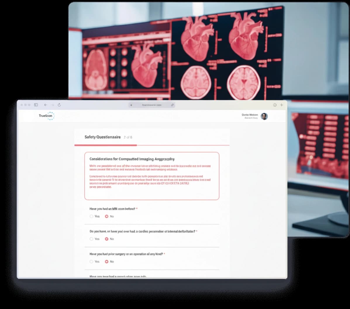
Complete Forms
Share important details such as:
- Pregnancy status
- Allergies to iodine or IV contrast
- Medications you take (including metformin)
Adjust Medications
- Stop stimulant meds (ADHD/energy/weight loss) for 36 hours
- Avoid erectile dysfunction meds (Viagra, Cialis, Levitra) for 72 hours
Day of Preparation
Follow these steps on the day of your scan:
- Fast for 4 hours
- Drink 2-4 glasses of water in the morning
- Avoid caffeine and chocolate
- Avoid smoking, vaping, or nicotine products
- Avoid vigorous exercise
At Your Scan
Check In
Arrive 30 minutes early. If your heart rate is already in range, the entire visit may take as little as 30 minutes. If medication is needed to lower it, plan for up to 1.5 hours (the scan itself takes ~10 minutes).
Standard & Safe Preparation
If your heart rate needs to be lowered, you’ll receive oral metoprolol. An IV will be placed, EKG leads attached, and nitroglycerin is given to dilate your arteries.
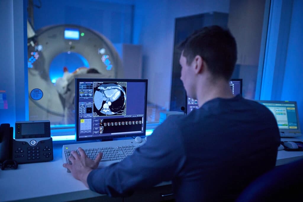
Imaging
We take initial pictures without contrast, then repeat with IV contrast. You may feel a brief warm sensation (1–3 minutes). Short breath holds are required to keep images clear.
Post-Imaging
Your IV is removed, vitals checked, and you’re free to leave shortly after.

After Your Scan
Stay Hydrated
Drink plenty of water to help your kidneys flush out the contrast dye.
Follow Guidelines
Avoid erectile dysfunction medications for 48 hours. Resume normal activities unless otherwise instructed.
Get Your Results
Within 7 days, you’ll receive a detailed report interpreted by a board-certified cardiologist. You’ll then have a video consultation with a TrueScan physician to review your results.
Frequently Asked Questions
To prepare safely and get the best images, follow these steps before your CCTA: • Forms: Complete your intake forms with details on pregnancy, allergies, and medications. • Fasting: Do not eat for 4 hours before your scan • Hydration: Drink 2 – 4 glasses of water the morning of your scan. • Caffeine/Chocolate: Avoid for on the day of your scan. • Nicotine: No smoking, vaping, or nicotine products on the day of your scan. • Exercise: Avoid vigorous physical activity on the day of your scan. • Medications: Hold blood pressure meds unless otherwise instructed. Stop stimulant meds (ADHD, energy, weight loss) for 36 hours. Avoid erectile dysfunction meds (Viagra, Cialis, Levitra) for 72 hours. If your physician ordered basic labs, including kidney function (Creatinine/GFR), and we haven’t received them yet, our team will contact you before your scan.
The AI Heart Scan itself only takes about 10 minutes, but your total visit may last 30 minutes to 1.5 hours. If your heart rate is already in range, you'll be scanned right away; if not, you may need extra time for medication to prepare you for the clearest images.
You'll lie on a padded table that moves through a wide, donut-shaped CT scanner (70 cm opening). A technologist will position you, place small EKG stickers on your chest to time images with your heartbeat, and start a small IV in your arm. • Standard prep in the room: Just before imaging, we'll place a nitroglycerin tablet under your tongue to gently widen the coronary arteries. When your heart rate is in range, we'll begin. • How the imaging works: We take a brief set of pictures without contrast, then inject IV contrast to make your arteries visible. You may feel a warm flush or a metallic taste for 1–3 minutes—both are normal and pass quickly. • Breath-holds: You'll hear clear instructions like 'Breathe in... hold.' Each breath-hold lasts about 5–10 seconds. Staying very still keeps the pictures sharp. • What you'll hear/feel: The scanner makes soft whirring sounds; it isn't loud like an MRI. The table moves smoothly through the ring—no tight tunnel. • Monitoring & communication: We continuously monitor your heart rhythm. You can speak with the technologist at any time through a two-way intercom, and we can pause if you need a moment. • Timing: The image acquisition itself takes about 10 minutes. When the images are complete, we remove the EKG stickers and IV, and you can head out shortly after.
Your AI Heart Scan report provides a complete, physician-interpreted overview of your coronary arteries. It includes: • Plaque analysis: Breakdown of plaque type (calcified, soft, and necrotic), plaque burden, and areas of stenosis (narrowing). • Blood flow assessment (FFR): Lesion-specific fractional flow reserve measurements showing whether plaque is restricting oxygen delivery to the heart. • Artery-by-artery detail: Visual maps of all major coronary vessels with AI quantification. • Cardiologist interpretation: Our board-certified cardiologist reviews your scan images and provides a diagnostic report. • Next steps: Our cardiologist typically includes recommendations for monitoring, lifestyle changes, or medications, along with information for your physician to guide care.
There are no universal guidelines yet, but the frequency generally depends on your risk level: • High-risk patients: every 1 year • Moderate-risk patients: every 2 years • Low-risk patients: every 3–5 years Your physician will determine the best interval for you based on your individual health profile.