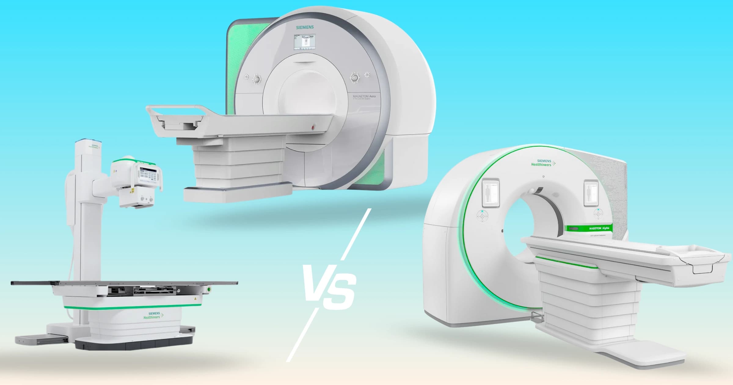
In a world where we rank cameras by megapixels and cars by horsepower, it is easy to want to look for a “best” imaging modality for your diagnostic appointment and be done with it. Surely there's a clearest, sharpest, most powerful scan that puts the rest to shame? One you'd always want to pick if time and money is no issue?
Surprise! (Or not really, since this article exists…) But medical imaging doesn't work that way. There is no single best scan. There is only the best scan for a specific question about a specific tissue: ultrasound, MRI, and CT don't compete so much as specialize.
So let's challenge the “best scan” myth head-on: starting with the modality that quietly does more than almost anyone gives it credit for.
Ultrasound: The Safe, Reliable First-Line
Ultrasound rarely makes the mental highlight reel. It's the scan associated with pregnancy, cold gel, and a fan-shaped grainy black-and-white image. Unglamorous at best.
But surprisingly if you had to design the ideal first-line imaging tool from scratch, you might well end up reinventing ultrasound.
What it is. Ultrasound works on the same principle as a bat: echolocation. A handheld transducer sends high-frequency sound waves far above the range of human hearing into the body.
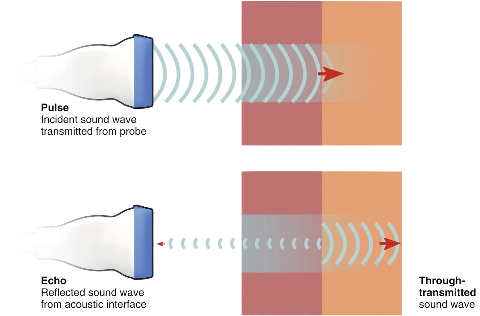
Image 1: Illustration of the mechanisms of ultrasound.
When those waves hit a boundary between two different tissues (i.e. the edge of the liver, or the wall of a blood vessel), some of the sound bounces back. The transducer listens for those echoes, measures how long they take to return and how strong they are, and a computer reconstructs a live, moving image from that information.
Why it's special. Ultrasound uses no ionizing radiation whatsoever. It's just sound. It's the reason it's the go-to for imaging a developing fetus, and the same safety profile means it can be used on nearly anyone: children, pregnant patients, people who need repeat scans month after month. There's no known ceiling on how many ultrasounds a person can safely have.
That safety unlocks a second superpower: repeatability. Because there's no dose to worry about and no lengthy setup, a clinician can scan you today, again tomorrow, and again the day after to watch how something changes.
More interestingly, ultrasound is one of the only modalities that shows the body moving in real time. A radiologist can watch a heart valve flap open and shut, watch blood surge through an artery, or press on a lump to see whether it's solid or fluid-filled. It's less like a photograph and more like a video call with your body.
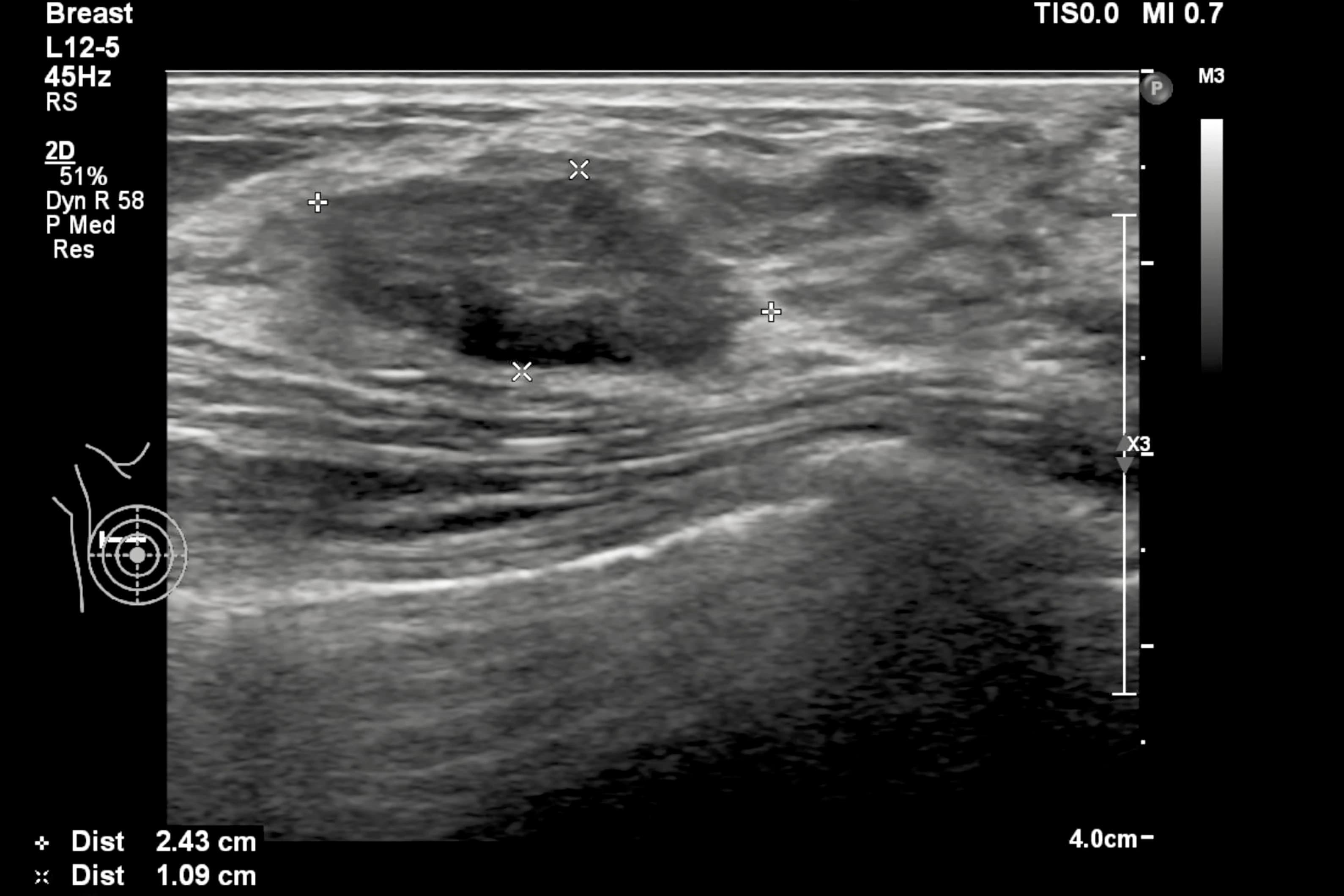
Image 2: Soft-tissue ultrasound of the breast.
Add to that its other virtues: it's typically the most affordable of the three, it's portable enough to bring to a hospital bedside, and it requires no needles, no contrast dye, and no enclosed tube (a necessity for anyone who feels uneasy in tight spaces).
What it's best for. Ultrasound excels at soft, fluid-rich, moving structures near the surface: the thyroid, gallbladder and bile ducts, the liver and kidneys, the ovaries and uterus, the testes, the breast, and the muscles and tendons. It's often the first stop for investigating a palpable lump, checking an organ, or just gaining abdominal insight.
The coolest quality of an ultrasound by far is its use of the Doppler effect (the law behind an ambulance siren changing pitch as it races past you). Ultrasound can measure the speed and direction of blood flow by listening to how the frequency of the returning echoes shifts as they bounce off moving red blood cells, it can detect a narrowed artery, a clot, or sluggish circulation, and render it on the screen.
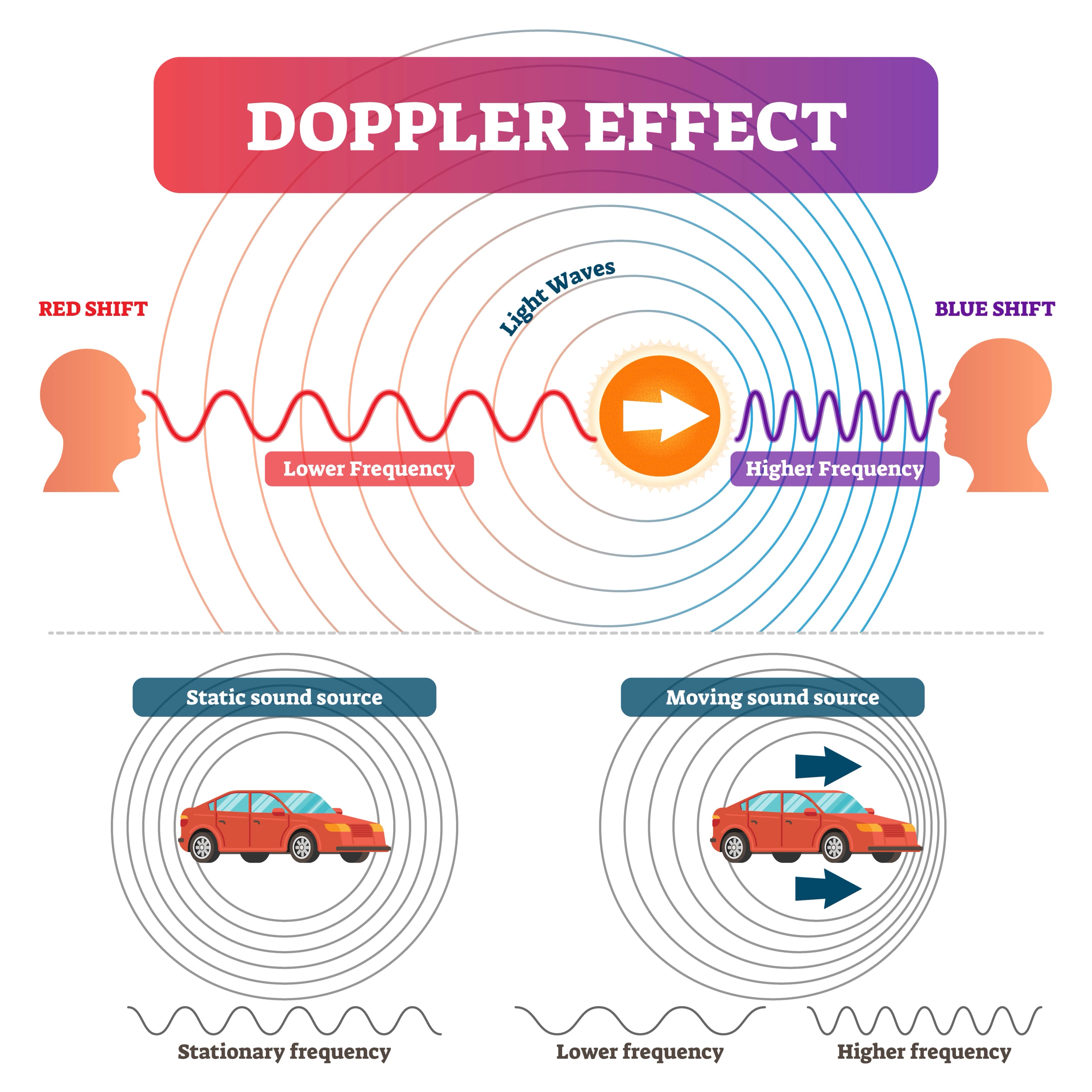
Image 3: Illustration of the Doppler effect.
Where it falls short. For all its power, sound waves have two enemies: air and bone. They scatter in gas and bounce off dense bone, so ultrasound struggles to see through the lungs, into the bowel, or inside the skull of an adult. It's also famously operator-dependent: the quality of the image leans heavily on the skill of the person holding the probe. And it can't peer deep into the body with the crisp, comprehensive detail that its two larger counterparts, the CT & the MRI, can provide.
Which raises the obvious question: if ultrasound is so good at watching blood flow, why is CT the scan doctors reach for when they need a definitive map of the body's vasculature?
CT: Speed and Density Specialist
What it is. A CT (computed tomography) scanner is, at heart, a very sophisticated X-ray machine. But instead of taking a single flat X-ray, it spins an X-ray source around your body, capturing hundreds of images from every angle in seconds. A computer then stacks those cross-sections into a detailed, three-dimensional map. Comparable to slicing a loaf of bread and being able to inspect every slice in every direction, and reassemble the whole loaf and rotate it freely.
CT's core skill is measuring density. X-rays pass easily through soft tissue (everything that's not bone or teeth) but are absorbed by dense material, which is why bone shows up bright white and air shows up black. CT reads these differences with exquisite precision.
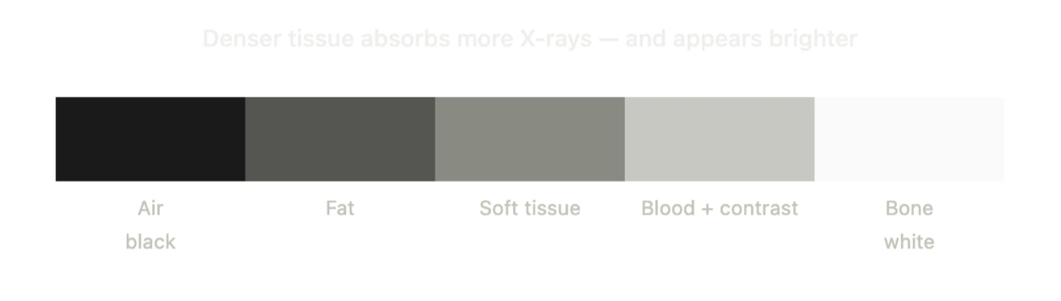
Image 4: Physiological color key for CT.
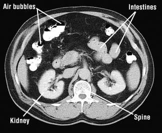
Image 5: CT cross-section illustrating the different values of organs depending on density.
Why it's amazing at vascular imaging. Two reasons that work in synchrony: first, speed. A modern CT can capture the entire chest or abdomen in the time it takes to hold a single breath. Blood is constantly moving and the heart never stops, so freezing that motion demands a fast shutter. It dominates in emergencies where seconds count, such as trauma, stroke, suspected internal bleeding.
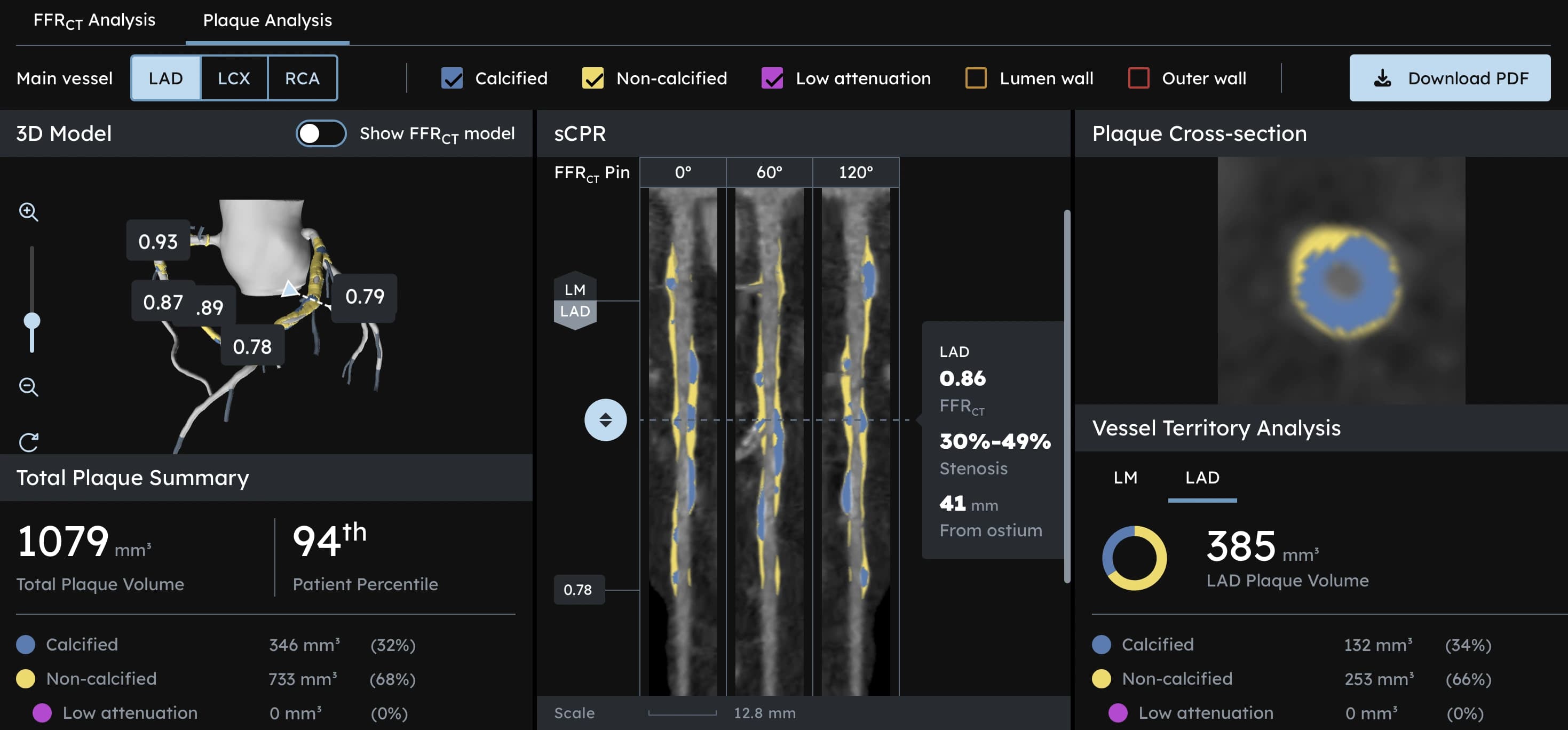
Second, and crucially, contrast. For vascular imaging (a CT angiogram, abbreviated as CTA), a patient is injected with an iodine-based contrast dye that lights up brilliantly under X-ray because iodine is extremely dense. As that dye courses through the arteries and veins, the fast-spinning scanner captures it in transit, producing an astonishingly crisp, complete road map of the vascular tree: every branch, blockage, aneurysm, or narrowing is rendered in high resolution. Ultrasound can tell you if blood is flowing well or sluggishly, but a CTA can produce a precise 3D blueprint of the entire arterial system before an operation. The combination of blistering speed and crisp contrast is exactly what vascular questions demand, and it's why CT owns that territory.
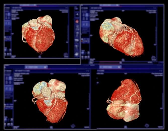
Image 6: CT Coronary Angiogram.
What else it's best for. Beyond blood vessels, CT is the master of bone, the lungs, and acute abdominal problems. Because it reads density so well, it's superb for detecting fractures, examining the air-filled lung, spotting kidney stones, and giving a fast, comprehensive survey of a badly injured or acutely ill patient. When a doctor needs a lot of accurate information about a lot of the body right now, CT is usually the answer.
Where it falls short. CT uses ionizing radiation so it isn't something to undergo on repeat, and it's used more cautiously in children and pregnancy. And while it's brilliant at density differences, it's comparatively weak at distinguishing between soft tissues that have similar densities. Ask a CT scan to tell you exactly where a tumor ends and healthy brain begins, or to grade the subtle damage inside a torn knee ligament, and it starts to squint…
For that kind of question, you need a machine that ignores density entirely.
MRI: The Soft-Tissue Expert
What it is. MRI (magnetic resonance imaging) is the most conceptually alien of the three, and maybe the most astonishing. It uses no radiation. Instead, it exploits the fact that your body is mostly water, and that every water molecule contains hydrogen atoms, which behave like tiny spinning magnets.
Slide into an MRI scanner and you enter an extraordinarily powerful magnetic field, one that coaxes all your hydrogen “compass needles” into alignment. The machine then pings them with a pulse of radio waves, knocking them briefly out of alignment. As they relax back into place, they all emit faint radio signals of their own. But hydrogen atoms in different tissues relax at different rates, and that's what gets measured. Hydrogen in fat behaves differently from hydrogen in muscle, which behaves differently from hydrogen in a tumor, or in inflamed tissue, or in the gray matter of the brain. The scanner listens to these subtle timing differences and builds an image of staggering soft-tissue contrast.
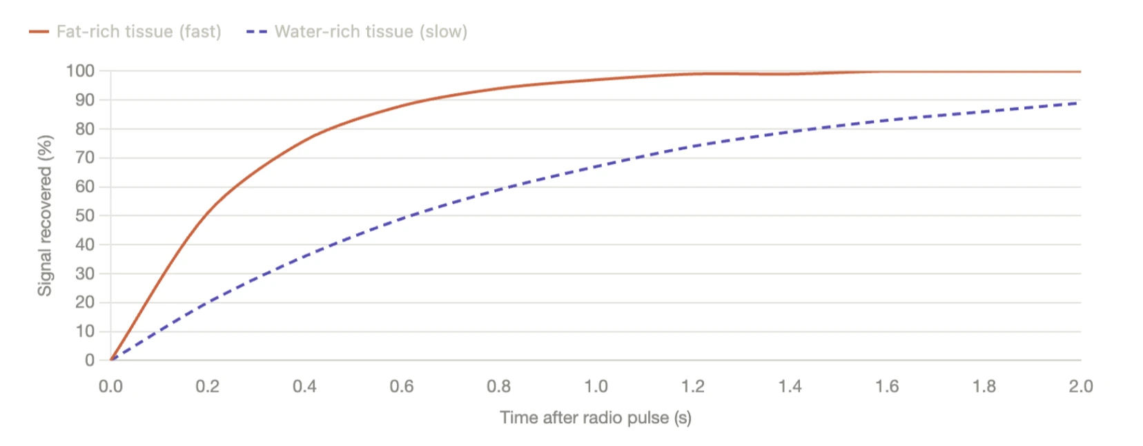
Image 7: Signal recovery of fat-rich tissue compared to water-rich tissue in MRI.
Why it rules soft tissue imaging. CT sees the body as a landscape of densities, but most soft tissues (muscle, ligament, nerve, and tumor) are similar in density. MRI ignores density completely and reads the molecular environment of the tissue: how water behaves inside it, how tightly it's bound, how quickly its hydrogen relaxes. Because that molecular behavior varies enormously between healthy and diseased soft tissue, MRI can pull them apart with amazing clarity.
The result is unmatched detail in exactly all the places CT struggles (and vice versa): the brain and spinal cord, the muscles, ligaments, tendons, and cartilage of joints, and the fine architecture of soft organs. It's why a torn knee ligament, a slipped spinal disc, a subtle brain lesion, an early-stage soft-tissue tumor, or the plaques of multiple sclerosis are MRI's home turf.
What it's best for. Neurological imaging (brain and spine), musculoskeletal injuries (joints, ligaments, cartilage), and detailed characterization of soft-tissue and organ abnormalities where telling one tissue type from another is the entire diagnostic challenge.
Where it falls short. MRI is slow. A detailed scan can take 30 to 60 minutes, and the patient must stay still throughout, which makes it poorly suited to emergencies or to imaging a beating heart without special techniques. It's the loudest and most claustrophobic of the three, taking place inside a long, narrow, clanging bore. The powerful magnet rules it out for some people with certain metal implants or devices. And it's typically the most expensive due to set-up and the technology.
So How Do You Actually Choose?
So an ultrasound listens, a CT measures density at high speed, and an MRI reads the molecular character of tissue. So how do you actually choose?
A few plain-language rules of thumb capture most of it:
- If it's about soft tissue, the brain, the spine, or a joint (and there is time to be thorough): That's MRI's world: unmatched detail, no radiation, but slow and enclosed.
- If it's urgent, or about bone, lungs, or blood vessels: That's CT: fast, comprehensive, and the definitive vascular mapper, at the cost of radiation exposure.
- If it's about a soft organ near the surface (ie anything abdominal), blood flow, a lump you can feel, or anything requiring real-time or repeated imaging, especially in a child, a pregnancy, or someone who'll need frequent follow-ups: Start with ultrasound: safe, live, affordable, and radiation-free.
In a side-to-side comparison, and in my own experience, ultrasound has been the go-to option. With no radiation, lengthy set-up, claustrophobia, dye, and is quiet, in addition to it being repeatable, it's frequently the first choice. In a great many cases it answers the question outright.
But there is no single best scan. Each tool is purpose-built and optimized for the question you hope to answer. So the next time you are at imaging crossroads, ask the useful question: what exactly are we trying to see?