
Heart disease has ruled as the #1 cause of death in America for over a century.
Every 40 seconds, someone in the U.S. has a heart attack – that's about 805,000 people every year.
How can people with completely different lifestyles share the same risk?
And how are there actually five different types of heart attacks?
The Five Types of Heart Attacks: The Quick Rundown
Medicine recognizes five distinct types of myocardial infarction (MI) – each with different causes, symptoms, and outcomes. Understanding the difference could literally save your life.
Type 1: The Classic Blockage (55% of cases)
The heart attack most people picture. A fatty plaque inside a coronary artery ruptures, triggering a blood clot that partially (NSTEMI) or completely (STEMI) blocks blood flow to the heart.
Type 2: The Supply-Demand Mismatch (32% of cases)
Here, there's no blockage – just an imbalance. The heart needs more oxygen than it's getting, often during severe illness, surgery, anemia, or extreme stress. Think of it as running your car engine in the red zone until it overheats.
Type 3: Sudden Cardiac Death
Some people die suddenly from what appears to be a heart attack before doctors can confirm it with blood tests. These patients often show signs of heart distress – like chest pain or changes on an EKG – but pass away before lab results (like troponin) can be taken or rise high enough to confirm a heart attack.
If someone has clear symptoms or EKG changes that suggest a heart attack, doctors classify it as a fatal heart attack even without blood test confirmation.
Types 4 & 5: Procedure-Related Heart Attacks
These occur during or after cardiac interventions.
- Type 4: Heart attacks linked to angioplasty or stent placement.
- Type 5: Heart attacks associated with coronary artery bypass surgery (CABG).
Type 1 Heart Attacks: The Classic Artery Blockage
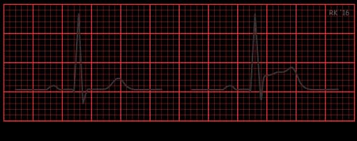
Image credit: cvphysiology.com
Myocardial infarctions (MIs) can also be broken down by whether the ST – the normally flat part of an ECG graph – is elevated or not.
STEMI: The Complete Blockage
In STEMI (ST-Elevation Myocardial Infarction), complete arterial blockage occurs. When this happens, blood flow stops instantly, and the heart muscle beyond the blockage begins to die within minutes.
What Causes STEMI?
Over years, cholesterol, inflammation, and plaque accumulate inside the arteries, forming unstable "vulnerable plaques." When one ruptures, it can trigger a massive blood clot that blocks blood flow – a process known as atherothrombosis.
Risk factors that make STEMI more likely include:
- High blood pressure
- High cholesterol
- Smoking
- Diabetes
Early Detection of STEMI
Advanced cardiac CT angiography (CTA) can spot these ticking time bombs before they rupture. The scan creates detailed 3D images of your coronary arteries, showing both the amount and type of plaque – including soft, high-risk buildup most likely to cause a heart attack.
Studies show that using CCTA proactively reduces heart attacks and deaths by 41% within just five years compared to standard care.
Treatment of STEMI
If a complete blockage is found, your cardiologist can restore blood flow by performing a stent procedure (PCI) or a coronary artery bypass surgery (CABG) to open the artery and reestablish circulation.
NSTEMI: The Partial Blockage
NSTEMI (Non–ST-Elevation Myocardial Infarction) occurs when a coronary artery is only partially blocked or blood flow is temporarily reduced – more of a traffic jam than a full roadblock. Unlike a STEMI, blood still reaches part of the heart muscle, so the injury is often less immediate but still serious.
What Causes NSTEMI?
NSTEMI is caused by the same plaque and inflammation process as STEMI, but the rupture or narrowing doesn't completely stop blood flow. Over time, this partial obstruction causes ongoing oxygen deprivation that can weaken the heart muscle.
Early Detection of NSTEMI
CCTA (Cardiac CT Angiography) is ideal for screening and identifying plaque buildup before it becomes dangerous. Once a heart attack occurs, Cardiac MRI is the gold standard for assessing heart muscle damage, scarring, and function – helping guide long-term treatment.
Treatment of NSTEMI
NSTEMI is less urgent than STEMI but still requires prompt care, including medications to stabilize plaque and cardiac catheterization to assess blockages and determine if stents or other interventions are needed.
Type 2 Heart Attacks: The Supply-Demand Mismatch
Type 2 myocardial infarction (T2MI) makes up about one-third of all heart attacks, yet most people have never heard of it. Unlike the classic Type 1 heart attack—caused by a ruptured plaque and blood clot—Type 2 happens when the heart's oxygen demand exceeds its supply. In other words, the heart is overworked.
Common Triggers
- Severe infections or fast heart rhythms (tachyarrhythmias) – involved in 55% of cases
- Major surgery or trauma
- Severe anemia or bleeding — which carries up to 47% mortality
- Extreme blood pressure (either very high or very low)
- Emotional or physical stress
Surprisingly, Type 2 heart attacks carry a higher long-term mortality rate than Type 1 – about 34% of patients die within 10 years, compared to 12% for Type 1.
How It's Detected
Diagnosis involves a mix of clinical evaluation, ECG changes, and elevated cardiac troponin levels – the key marker of heart muscle injury. The goal is to confirm that ischemia (reduced blood flow) is the cause, even when no clot or blockage is present.
Special Forms of Type 2 Heart Attacks
Type 2 heart attacks can appear in several unique forms that don't involve a classic blockage but still cause real heart injury. These include Broken Heart Syndrome – triggered by extreme emotional or physical stress – and Small Vessel Disease, which affects the tiniest coronary arteries.
Broken Heart Syndrome
Takotsubo Cardiomyopathy is a fascinating – and frightening – manifestation of T2MI. Also known as "broken heart syndrome," it mostly affects older women and mimics a traditional heart attack. Triggered by sudden emotional or physical stress (like the loss of a loved one, a major argument, or even overwhelming joy), the heart's main pumping chamber balloons into an odd, "octopus trap" shape.
The cause is a surge of stress hormones (catecholamines) that stuns the heart muscle. Cardiac MRI confirms the diagnosis by showing the ballooning pattern without any arterial blockages.
Small Vessel Disease (Microvascular Angina)
Another subtle form of Type 2 heart attack, small vessel disease affects the tiniest coronary arteries – vessels no wider than a few human hairs. It predominantly affects women after menopause and often goes undiagnosed because standard angiograms look normal.
What happens:
Tiny vessels can't dilate properly during increased demand. It's like having functional traffic lights but perpetually narrowed lanes.
Detection:
Requires specialized testing like coronary flow reserve measurements or nuclear stress testing with PET scans. Advanced cardiac MRI can detect microvascular dysfunction that other tests miss, providing crucial early intervention opportunities.
Your Risk Profile: What You Can Control
Understanding your personal risk profile remains crucial for prevention. Many non-modifiable risks such as age, gender, genetics, and family history are factors to be taken into account, but modifiable risks are those that matter most.
Modifiable Risk Factors
- Smoking (strongest association with heart attacks)
- High cholesterol
- High blood pressure
- Diabetes
- Obesity
- Sedentary lifestyles and physical inactivity
- Poor diet high in saturated fats, trans fats, sodium, and added sugars
- Excessive alcohol consumption
Studies show that following 4+ healthy lifestyle factors reduces heart failure risk by 45%. Physical activity, maintaining normal weight, modest alcohol use, and not smoking provide the greatest protection.
The Prevention Revolution: Advanced Screening & AI
Modern Imaging Technology
Heart attack treatment has transformed dramatically over five decades. CCTA and MRI technology have revolutionized preventive cardiology. Modern scanners acquire images in a single heartbeat, revealing:
- Plaque composition and vulnerability
- Arterial narrowing before symptoms develop
- Risk stratification with extraordinary accuracy
People who undergo CCTA are far more likely to start preventive treatments early – when they make the biggest difference.
Preventive Medications That Save Lives
These medications can protect arteries, stabilize plaque, and prevent future heart attacks:
 Dual antiplatelet therapy (aspirin plus P2Y12 inhibitors) to prevent clots
- Statins to lower cholesterol and reduce inflammation
- ACE inhibitors to protect the heart and improve circulation
- Beta-blockers to reduce heart strain
- Advanced anticoagulants to lower the risk of dangerous blood clots
The AI Breakthrough
AI represents the next great leap in preventing heart attacks.
Through the analysis of medical imaging (like CCTA, CT, and ECGs), AI algorithms can detect subtle signs of plaque buildup, vessel inflammation, and blood-flow changes that even experienced clinicians might miss. These models analyze millions of data points across large patient populations, recognizing complex patterns that signal risk long before traditional testing does.
This precision allows physicians to assess cardiovascular risk years in advance – not after a heart attack, but while the disease is still reversible. Patients can start targeted preventive therapies earlier, from cholesterol-lowering statins to lifestyle interventions that stabilize plaque and restore blood flow.
This remarkable achievement reflects advances in emergency care, interventional cardiology, and preventive medicine.
The results speak for themselves: since 1970, heart attack mortality has plummeted by over 90%, thanks to advances in emergency response, interventional cardiology, and preventive care. AI now extends that progress even further – shifting medicine from reacting to heart attacks after they happen to predicting and preventing them before they begin.
The Bottom Line: Survival & Prevention
The results speak for themselves: since 1970, heart attack mortality has plummeted by over 90%, thanks to advances in emergency response, interventional cardiology, and preventive care. AI now extends that progress even further – shifting medicine from reacting to heart attacks after they happen to predicting and preventing them before they begin.
What This Means for You
Heart attacks aren't a single condition – they're a spectrum of diseases with different causes, presentations, and outcomes. Survival today isn't just about getting to the hospital in time – it's about finding the problem early enough that you never end up there at all.
With advanced imaging like our AI Heart Scan, we can now see inside the heart with stunning precision – spotting dangerous plaque and restricted blood flow years before symptoms appear.
These breakthroughs give you the chance to detect and reverse dangerous plaque before it causes harm. When you know early, you can act early – protecting your arteries, improving heart function, and even undoing years of silent buildup.
Learn more about how to aggressively treat and stabilize soft plaque in our follow-up article: How to Aggressively Treat Dangerous Soft Plaque.
Your Action Plan
Your best defense? Know your risk and get screened proactively. Because the greatest victory over heart disease is not surviving it – it's never letting it happen in the first place.
References
[1] Abdelrahman, K. M., Nasir, K., & Blaha, M. J. (2020). Coronary computed tomography angiography from clinical uses to emerging technologies. Journal of the American College of Cardiology, 76(10), 1226–1243. link
[2] Ahmad, S. A., Brito, D., & Garlapati, P. (2023). Takotsubo cardiomyopathy. In StatPearls. StatPearls Publishing. PMID: 29489280 link
[3] Akbar, H., Foth, C., & Kahloon, R. A. (2024). Acute ST-segment elevation myocardial infarction (STEMI). In StatPearls. StatPearls Publishing. PMID: 29489224 link
[4] Almansouri, N. E., Almansour, F. T., & Albattat, M. H. (2024). Early diagnosis of cardiovascular diseases in the era of artificial intelligence. Journal of Personalized Medicine, 14(3), 282. link
[5] American Heart Association. (2020). Incidence, trends, and outcomes of type 2 myocardial infarction. Circulation, 141(7), 549–558. link
[6] American Heart Association. (2023). Artificial intelligence may speed heart attack diagnosis and treatment. AHA Scientific Sessions. link
[7] American Heart Association. (2024). 2024 heart disease and stroke statistics update fact sheet. AHA Statistical Update. link
[8] American Heart Association. (2024). Cardiac computed tomography angiography (CCTA). AHA Patient Education. link
[9] Basit, H., Malik, A., & Huecker, M. R. (2023). Non–ST-segment elevation myocardial infarction. In StatPearls. StatPearls Publishing. PMID: 29494106 link
[10] British Heart Foundation. (2024). What are the different types of heart attacks. BHF Heart Matters. link
[11] Bularga, A., Lee, K. K., Stewart, S., Ferry, A. V., Chapman, A. R., Marshall, L., ... Mills, N. L. (2022). Assessment of oxygen supply-demand imbalance and outcomes in patients with type 2 myocardial infarction. JAMA Network Open, 5(6), e2218162. link
[12] Del Gobbo, L. C., Kalantarian, S., de Boer, I. H., Kizer, J. R., Biggs, M. L., Ix, J. H., ... Djoussé, L. (2015). Contribution of major lifestyle risk factors for incident heart failure in older adults. Journal of the American College of Cardiology, 66(20), 2263–2274. link
[13] Harvard Health Publishing. (2023). Broken-heart syndrome (takotsubo cardiomyopathy). Harvard Health Publishing. link
[14] Kosaraju, A., Goyal, A., & Grigorova, Y. (2023). Cardiogenic shock. In StatPearls. StatPearls Publishing. PMID: 29494020
[15] Martin, S. S., Blumenthal, R. S., Miller, M., Michos, E. D., Jones, S. R., & Virani, S. S. (2024). 2024 heart disease and stroke statistics: A report of US and global data from the American Heart Association. Circulation, 149(8), e347–e913. link
[16] Mayo Clinic. (2021). Small vessel disease – Symptoms & causes. Mayo Clinic Health Information. link
[17] Ramjattan, N. A., Cheema, M., & Balla, S. (2024). Coronary CT angiography. In StatPearls. StatPearls Publishing. PMID: 29494068 link
[18] Rippe, J. M. (2018). Lifestyle strategies for risk factor reduction, prevention, and treatment of cardiovascular disease. American Journal of Lifestyle Medicine, 13(2), 204–212. link
[19] Sandoval, Y., Smith, S. W., & Apple, F. S. (2014). Supply/demand type 2 myocardial infarction: Should we be paying more attention? Journal of the American College of Cardiology, 63(20), 2079–2087. link
[20] SCIRP. (2018). Myocardial infarction: An overview of STEMI and NSTEMI. Scientific Research Publishing. link
[21] Thygesen, K., Alpert, J. S., Jaffe, A. S., Chaitman, B. R., Bax, J. J., Morrow, D. A., & White, H. D. (2018). Fourth universal definition of myocardial infarction. Circulation, 138(20), e618–e651. link
[22] UT Southwestern Medical Center. (2023). Broken heart syndrome (takotsubo cardiomyopathy). UT Southwestern Cardiology. link
[23] Wereski, R., Kimenai, D. M., Taggart, C., Cartwright, N., Lowe, D. J., ... Mills, N. L. (2022). Risk factors for type 1 and type 2 myocardial infarction. European Heart Journal, 43(2), 127–135. link


